Cleft & Craniofacial Surgery
OMS and Other Dental Specialists
Session: Oral Abstract Session Two
SA2 - The Impact of Premature Fusion Intersphenoid Synchondrosis (ISS) in Costello Syndrome
Friday, September 13, 2024
1:45 PM - 1:55 PM EDT
Location: W315B, Orange County Convention Center

Susan Keefe, DDS
Resident
University of North Carolina
Durham, North Carolina
Oral Abstract Presenter(s)
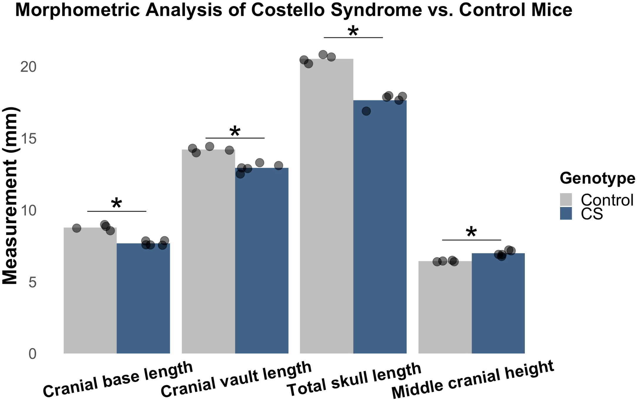
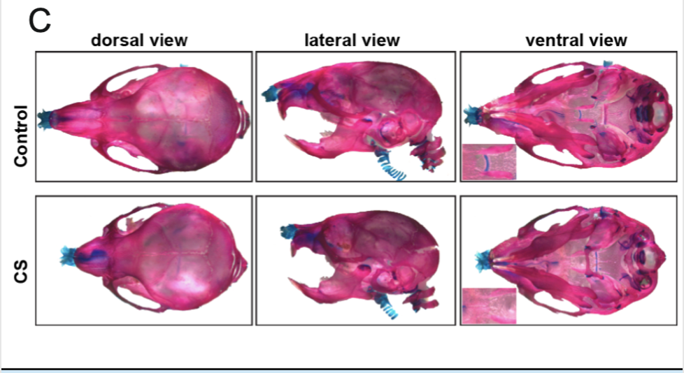
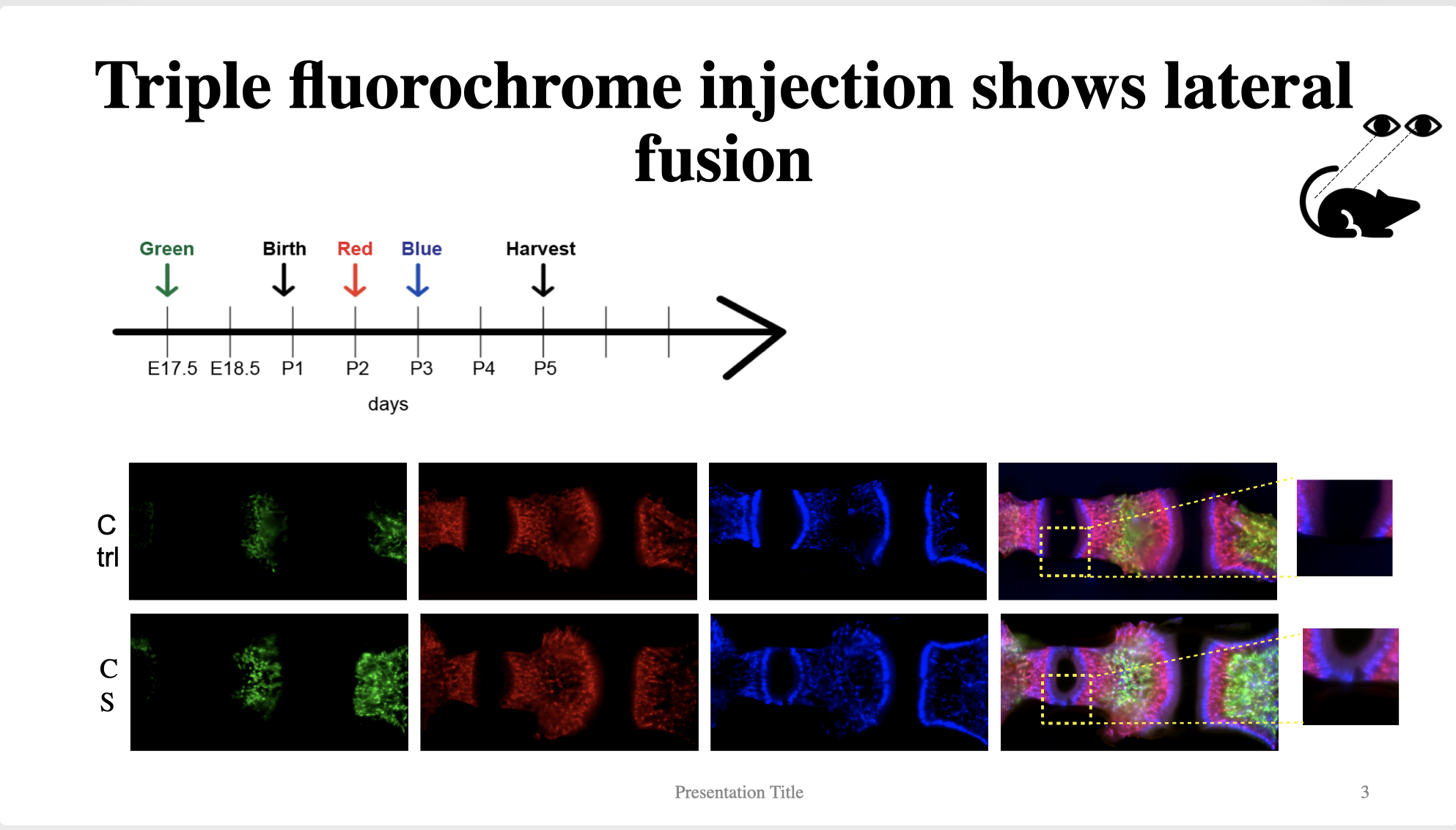
Abstract: Costello Syndrome (CS) is caused by heterozygous de novo germ line mutations in HRAS that lead to Ras/MAPK dysregulation. CS patients often manifest numerous extracranial anomalies affecting the cardiac, nervous, and musculoskeletal systems, alongside dysmorphic craniofacial characteristics such as a prominent forehead, distinct mid-facial features, and various forms of tooth malocclusions. Though, the mechanisms underlying craniofacial manifestations of the syndrome is not clear. In a CS mouse model, we observed abnormal development of the intersphenoid synchondrosis (ISS), one of the main growth centers responsible for longitudinal growth of the cranial base. This discovery suggests a potential correlation between facial development and the growth of cranial base synchondrosis. The aim of this study is to investigate the fundamental developmental mechanisms contributing to craniofacial abnormalities in CS associated with the cranial base. Postnatal-day 21 mouse skulls were analyzed by micro-C T and skeletal preparation. Unpaired student t test was used to compare the means of control and CS mouse skull morphogenetic analysis parameters, such as maxilla length, cranial base length, carnival vault length, middle cranial height, and total skull length. Fluorochrome injections were used to assess ISS mineralization patterns. Lastly, ISS explants were cultured using Trowell’s method.CS mice exhibit craniofacial abnormalities similar to those seen in CS patients such asshorter cranial base (p = 0.0013), maxilla (p = 0.00043), cranial vault (p = 0.025), total skull length (p= 0.000015) and greater middle cranial height (p = 0.0004) compared to control mice. Skeletal preparations of P21 mice show a distinct skull shape in CS mice and an absence of the cartilaginous portion of ISS in CS mice. No difference in utero bone formation is observed. The start of bridging of the ISS in CS occurs around post-natal day 3 and is initiated at the lateral edges of the synchondrosis by abnormal chondrocyte differentiation. Explants of the ISS show the fusion does not continue in vivo. This study shows that abnormal CS mouse craniofacial development originates from the fusion of ISS. The lateral fusion of the ISS first occurs at P3 and is related to the ectopic expression of bone in a noncell-autonomous fashion. The importance of other growth centers such as spheno-occipital synchondrosis which fuses much later in humans should also be investigated in its role in craniofacial development in adolescents. 1. Goodwin et al., Am J Med Genet A, 2014. 2. Cao et al., Ortho Craniofac Res, 2017.3. Schuhmacher et al., JCI, 2008.
morphogenic analysis
skeletal prep of mouse skull
fluorochrome injection
morphogenic analysis

skeletal prep of mouse skull

fluorochrome injection


